Vascular testing for rheumatology is the use of non-invasive arterial and autonomic studies, principally the ankle-brachial index (ABI), the toe-brachial index (TBI), volume plethysmography waveforms, sudomotor testing and the cardiac autonomic reflex battery, to put an objective number on the circulatory and autonomic status of patients whose inflammatory disease already places them in a higher cardiovascular risk category. It belongs in a rheumatology practice for a narrow and practical reason: the population is unusually rich in guideline-recognized cardiovascular risk, and unusually poor in reliable physical findings.
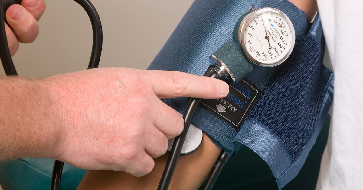
The 2018 AHA/ACC multisociety cholesterol guideline names chronic inflammatory disorders such as rheumatoid arthritis and psoriasis as risk-enhancing factors, and it names an ankle-brachial index below 0.9 as a separate risk-enhancing factor in the same list. A rheumatology panel therefore arrives already carrying one item off that list, and the measurement that would identify a second is a cuff-based study that takes roughly 7 to 10 minutes in an exam room.
This guide covers what the studies measure, why the rheumatology population is different, which patients warrant testing, when the ABI cannot be trusted, what the autonomic portion adds, which patients the equipment cannot be used on, and how a practice brings the capability in-house. It is written for physicians and practice administrators, and it stays deliberately close to what the equipment documentation and the published guidelines actually say.
What does vascular testing for rheumatology actually measure?
Less than most clinicians expect, and none of it invasive. A point-of-care platform measures systolic pressure at both ankles and both arms to produce the ankle-brachial index and its normal reference ranges, measures toe pressure to produce the toe-brachial index, records volume plethysmography waveforms that describe the shape of arterial flow rather than a single number, and performs sudomotor testing of the sympathetic skin response.
Alongside those arterial measures sits the autonomic portion, and it is worth describing precisely because it is routinely assumed to be a passive heart-rate-variability recording. On the TM-Flow platform that HealthWright Technologies supplies as CMAT Advantage, the manufacturer’s instructions for use document three provoked maneuvers: sixty seconds of paced deep breathing at five seconds in and five seconds out to derive the expiration-to-inspiration ratio, a Valsalva maneuver performed against a manometer at 40 mmHg for fifteen seconds to derive the Valsalva ratio, and a standing maneuver that captures the 30:15 heart-rate ratio together with the systolic and diastolic blood-pressure response to standing. The sequence runs Valsalva, then deep breathing, then stand.
The systems are built on FDA-cleared component devices, with clearance held by the manufacturer, LD Technology. The equipment is on-premises. The study runs in the office, the report is produced on the system in the office, and it is printed or saved to the chart during the same visit.

Why does a rheumatology panel carry more cardiovascular risk than the chart suggests?
Because standard risk calculators were not built on this population, and the specialty societies have said so for well over a decade.
EULAR’s 2015/2016 update on cardiovascular disease risk management in inflammatory joint disorders sets out three positions that matter operationally. Cardiovascular risk is higher in rheumatoid arthritis and may also be higher in ankylosing spondylitis and psoriatic arthritis. Comprehensive risk assessment should be performed at least once every five years, and reconsidered after major changes in antirheumatic therapy. And risk prediction models should be adapted for rheumatoid arthritis by a 1.5 multiplication factor when disease-specific terms are not already built into the model. The task force also placed responsibility for cardiovascular risk management with the rheumatologist rather than assuming primary care will absorb it.
Two consequences follow for a practice. The first is that a five-year reassessment interval is a floor, not a ceiling, and it lands inside a specialty that already sees these patients several times a year. The second is that a risk model adjusted upward by a fixed multiplier is an estimate, and an estimate is not a measurement. Vascular testing for rheumatology replaces one of the estimated inputs with a measured one.
None of this means every rheumatology patient should be tested. It means the population justifies the capability, and the individual patient still has to justify the study.
Which rheumatology patients warrant vascular testing?
The 2024 ACC/AHA multisociety lower extremity peripheral artery disease guideline is direct about the trigger. In patients with a history or physical examination findings suggestive of peripheral artery disease, a resting ankle-brachial index is recommended to establish the diagnosis, a Class 1 recommendation. In patients at increased risk, a resting ABI is described as reasonable, at Class 2a. Worth noting for accuracy: that guideline lists its risk amplifiers explicitly, and inflammatory or autoimmune disease is not among them. The risk-enhancing status of rheumatoid arthritis comes from the cholesterol and primary prevention guidelines, not from the PAD guideline, and copy that blurs the two is overstating the case.
In practice the patients who generate a defensible indication in a rheumatology clinic tend to share a problem: a limb complaint that the joint findings do not fully explain. Calf pain attributed to deconditioning in a patient who has stopped walking far enough to provoke anything. A cool, discolored foot in a long-standing seropositive patient. Burning feet in a patient with sicca symptoms. Non-healing skin over a pressure point. Diminished or absent pedal pulses noted on an examination that was really looking at the ankle joint.
The discipline that separates appropriate testing from the conduct that gets practices into trouble is simple and it is not negotiable: the symptoms or examination findings are documented first, the study is ordered because of them, and the result informs what happens next. Testing an asymptomatic panel on a schedule is a different activity with a different regulatory profile, and this article is not an argument for it.
When can the ankle-brachial index not be trusted?
This question matters more in rheumatology than in most specialties. A normal resting ABI sits between 1.00 and 1.40. A value at or below 0.90 is the conventional threshold for peripheral artery disease. A value above 1.40 does not mean excellent circulation, it means the vessel would not compress, and the number is therefore uninterpretable.
Non-compressible vessels are common in exactly the comorbidities a rheumatology panel accumulates: long-standing diabetes, chronic kidney disease, and advanced age. The toe vessels are far less prone to medial calcification, which is why the toe-brachial index is the fallback measurement when the ankle number cannot be believed. A platform that captures toe pressure in the same sitting removes a second appointment from the pathway.
Waveform morphology is the third input. A volume plethysmography tracing that has lost its dicrotic notch and flattened tells you something a single ratio cannot, and it remains readable when the pressure numbers are compromised.
What does the autonomic portion of the study add?
A different axis of information. Arterial measurement describes conduit flow. The cardiac autonomic reflex battery describes how the nervous system regulates heart rate and blood pressure under provocation, and the sudomotor component describes sympathetic control of the sweat glands.
Two of those measures speak to symptoms rheumatology clinics hear constantly and rarely quantify. Lightheadedness on standing is usually attributed to medication, deconditioning or dehydration, and the standing maneuver records the actual systolic and diastolic response rather than the assumption. Distal burning, tingling and dryness are usually attributed to the disease or to a drug, and the sympathetic skin response records something measurable about small-fiber sympathetic function.
What the battery does not do is diagnose the underlying rheumatic disease, establish its activity, or replace serology and imaging. Final interpretation of the study is the sole responsibility of the practitioner, read against the case history, symptoms, known diagnoses, current medications and treatment plan. The manufacturer’s own documentation says exactly that, and it is the right posture.
Which rheumatology patients cannot have the full study?
This is the section most articles on in-office diagnostics leave out, and it is the one a rheumatology practice should read first, because this specialty concentrates several of the relevant conditions. The contraindications below come from LD Technology’s own contraindications and warnings document for the platform rather than from general clinical literature.
The single most rheumatology-specific item: metal pins or prostheses in the legs are a contraindication to the sudomotor component. A practice managing long-standing inflammatory arthritis carries an unusually high proportion of patients with knee and hip arthroplasty and other lower-limb hardware, so this will not be an occasional exception. Plantar dermatological lesions or calluses rule the sudomotor component out as well, which catches psoriatic plaques and neuropathic skin changes over pressure points.
On the pressure side, the cuff-based ABI and TBI component must not be used in the presence of an arterial catheter, an arteriovenous fistula or shunt, or a pressure dressing on the limb, and it must not be used where perfusion is low, because inflating a cuff over poorly perfused tissue risks skin erosion or pressure necrosis. That last item deserves emphasis in a specialty that sees critical digital and pedal ischemia: the patient whose circulation looks worst is not automatically the best candidate for a cuff study.
The Valsalva maneuver carries its own exclusions. It should not be performed in patients with proliferative retinopathy, in patients who have had laser treatment for retinopathy within the previous three months, or when systolic blood pressure is 160 mmHg or higher. The last of those is worth flagging for a rheumatology workflow, because patients on long-term corticosteroid therapy frequently present with elevated pressures, and a reading taken at the start of the visit determines whether that maneuver can proceed at all.
| Finding in the patient | Component affected | What the practice does |
|---|---|---|
| Metal pins or prostheses in the legs | Sudomotor | Run the arterial components only |
| Plantar lesions or calluses | Sudomotor | Run the arterial components only |
| AV fistula, arterial catheter or pressure dressing | Cuff-based ABI and TBI | Do not inflate on that limb |
| Low perfusion in the limb | Cuff-based ABI and TBI | Do not use; risk of skin erosion or necrosis |
| Systolic BP 160 mmHg or higher, or recent retinal laser | Valsalva maneuver | Omit that maneuver |
| Bilateral mastectomy, or absence of two arms or two legs | Cuff-based ABI and TBI | Study not appropriate |
Two operational details make the difference between a workable clinic day and a wasted appointment. First, a contraindication to one component does not cancel the visit: the measurement type is selected before the examination begins, so a patient with lower-limb hardware can still have the arterial study performed. Second, the sympathetic skin response habituates. Amplitude falls after repeated electrical stimulation, and the manufacturer advises against repeating that test in the same patient inside twenty minutes, which is a scheduling constraint rather than a clinical one but it will bite a practice that tries to re-run a study immediately.
How is vascular testing for rheumatology coded and documented?
Three code families cover the studies described here. The bilateral lower extremity arterial study with pressures and waveforms is reported under CPT 93923. Sudomotor testing falls under 95923. The cardiovagal battery falls under 95921, whose descriptor requires two or more of the deep-breathing heart-rate response, the Valsalva ratio and the 30:15 ratio, all three of which the platform documents performing.
Three points about coding that practices get wrong. The manufacturer does not assign CPT codes and does not purport to; its software instructs the interpreting physician to choose the relevant codes. Coverage is determined by your Medicare Administrative Contractor’s local coverage determination, which you should read rather than assume. And documentation of medical necessity is the load-bearing element, not the presence of the equipment: the indication, the findings and the effect on management all belong in the note. Published Medicare fee-schedule amounts for these codes are available in the CMS physician fee schedule and change every January.
On staffing, the study is performed by trained clinical personnel and interpreted by the physician. Supervision requirements and who may operate the equipment are covered in detail in our guide to who can perform an ABI test.
How does a rheumatology practice bring vascular testing in-house?
The practical requirements are modest and they are mostly about consistency rather than equipment. A quiet exam room where a patient can lie comfortably. Staff trained to position the cuffs correctly, because an incorrectly positioned applied part produces an inaccurate reading and no amount of downstream interpretation fixes that. A rule about when the study is not run, since pressures taken within an hour of a meal, after exercise, after coffee or alcohol, after a bath, or within an hour of smoking are unreliable. And a standing habit of taking the reading with the patient settled, because anxiety in the room moves the number.
The environment matters more than most buyers expect. The manufacturer warns against carpeted flooring and relative humidity below 30 percent, both of which raise electrostatic discharge, and against operating the wireless components within ten feet of a router, a cell phone or a microwave. A practice that installs the system in a carpeted back office next to the staff kitchen has created a support ticket rather than a service line.
For the wider view of how an in-office arterial and autonomic service line fits alongside existing workflow, see our overview of the in-office vascular testing and what it involves for independent practices, which serves as the hub for this series. Practices evaluating the equipment itself can review the TM-Flow ANS testing device that HealthWright supplies, or read what is included in the CMAT Advantage package.
Frequently asked questions about vascular testing for rheumatology
What is vascular testing for rheumatology?
Vascular testing for rheumatology is non-invasive, in-office measurement of arterial and autonomic function in patients with inflammatory rheumatic disease. The panel typically includes the ankle-brachial index, the toe-brachial index, volume plethysmography waveforms, sudomotor testing and a provoked cardiac autonomic reflex battery. It is performed when a documented symptom or examination finding raises the question, and the result informs clinical management.
Which rheumatology patients should have an ankle-brachial index?
The 2024 ACC/AHA lower extremity peripheral artery disease guideline recommends a resting ankle-brachial index to establish the diagnosis in any patient whose history or physical examination findings suggest peripheral artery disease, and describes a resting ABI as reasonable in patients at increased risk. In a rheumatology clinic that usually means the patient with a limb complaint, a skin change or a pulse finding that the joint disease does not fully explain.
Can a patient with a knee or hip replacement have the full study?
Not the sudomotor component. The manufacturer lists metal pins or prostheses in the legs as a contraindication for that device. The arterial components can still be performed, because the measurement type is selected before the examination starts. This matters in rheumatology, where lower-limb arthroplasty is common.
Which CPT codes apply to in-office vascular and autonomic testing?
CPT 93923 covers the bilateral lower extremity arterial study with pressures and waveforms. CPT 95923 covers sudomotor testing, and CPT 95921 covers the cardiovagal battery. The interpreting physician selects the codes, coverage is governed by the local coverage determination of your Medicare Administrative Contractor, and medical necessity must be documented in the note.
How long does the study take and who performs it?
Roughly 7 to 10 minutes on a point-of-care platform, performed by trained clinical staff in an exam room and interpreted by the physician. The report is generated on the system in the office and can be printed or saved to the chart during the same visit.
Bringing the capability into your practice
HealthWright Technologies has supplied diagnostic and therapeutic equipment to independent physicians since 2000, with training, implementation and ongoing support included rather than sold separately. If you are weighing whether vascular testing for rheumatology fits your patient mix and your exam-room capacity, we will walk through the measurements, the documentation expectations and the workflow with you before anything is quoted.
Reach us at our contact page, by email at contact@healthwrighttechnologies.com, or by phone at (678) 322-7146. HealthWright Technologies, 60 Bear Creek Marina Road, Mansfield, GA 30055.
This article is educational and is intended for clinicians and practice administrators. It is not medical advice and it is not a coding or coverage determination. Consult your healthcare provider before starting any new treatment. CPT is a registered trademark of the American Medical Association. CPT codes, descriptions and other data are copyright the American Medical Association; all rights reserved. Code selection, medical necessity and coverage remain the responsibility of the treating and billing provider, and coverage is governed by the applicable local coverage determination. Device contraindications summarized here are drawn from manufacturer documentation and should be confirmed against the instructions for use supplied with your system.
Ready to see the CMAT Advantage in your practice?
Book a free 25-minute demo: see the full PAD + autonomic workup and how the three established CPT codes apply, with no obligation. Backed by our 60-day buyback guarantee.
Book a Free Demo →